ASSIGNMENT 2: The respiratory System
TASK 1
AIM
The purpose of the experiment that we carried out was to investigate the effect that exercise has on our breathing rate. We created a short exercise program which was to be repeated a certain number of times for each test. For example the first task the program was done once without repetition, but during the second test the program was repeated twice. In total we tested how 6 periods of exercise with different lengths affected the breathing rate of someone doing exercise. Between each exercise period we measured the breathing rate by counting how many breaths per minute, using a stop watch to increase the accuracy of our measurements. From our results we wanted to find a clear pattern on how changing the length of exercise can affect the breathing rate.
INTRODUCTION
The respiratory system's main purpose is to provide Oxygen to the blood so it can be transported around the body. The respiratory system is made up of the nasal cavity, the two lungs, the throat and the larynx, bronchi and trachea. We wanted to investigate the effect that exercise has on our breathing rates. To do this we nominated one person to do the exercise and then created a short exercise plan which you can see below.

APPARATUS LIST
- Stop watch
- Pencil
- Paper
- Ruler
METHOD
1. Take the breathing rate for one minute at rest before any exercise has take place
2. Exercise for a total of 30 seconds, 60 seconds, 90 seconds, 120 seconds, 150 seconds and 180 seconds and measure the breathing rate for a minute between each length of exercise
3. take note of the results and plot on a graph
There are a few things that we had to consider before we carried out our investigation. These are independent variables, the dependent variables and the control variables of the experiment. We decided that the independent variables would be the length of exercise per test. We would change this by increasing the length of each move by ten seconds each time. Our dependent variables would be the breathing rate. We would measure the breathing rate for a minute between each exercise period using a stop watch and simply just counting how many breaths we taken in that minute. The control variables we decided upon were firstly the same person doing the exercise because everybody's fitness levels vary so using different people for each test wouldn't give us reliable results. This person should wear the same clothing throughout the investigation, the same room should be used for the exercise and finally the same stopwatch and the same person using the stop watch to minimize the chance of errors.
RESULTS
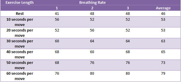
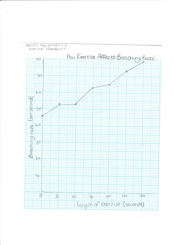
Our results show a definite trend. From the graph as well as the results table you can see that as the length of exercise increases so does the breathing rate. This is because the Oxygen in the body is being used up quicker to power the muscles used for exercise so the respiratory system has to work harder to provide enough oxygen to the body so it can still function.
There are a few anomalous results in our set of data also. At 30 seconds and 60 seconds the average result for the breathing rate is the same. This could be down to during one of the tests we could have left it too long to check the breathing rate or our way of measuring the breathing rate was not very accurate.
CONCLUSION
In conclusion, our investigation showed us that the longer that you exercise for, the higher your breathing rate will be. This is because the oxygen in your body is being used quicker so your body needs to replace it quicker than if you were at rest. Our results clearly support this even with the couple of anomalous results that we did acquire.
EVALUATION
A way this investigation could be improved would be to use a more accurate way of calculating the breathing rate than counting each breath per minute. A more accurate way to measure breathing rate would be to use a spirometer. A spirometer is much more accurate as you can set one rotation to last exactly one minute and it draws each breath directly on to a graph.
Another way that the investigation could be improved is measuring more than one persons breathing rate and seeing what the effect of exercise is on a different number of people and comparing the results. It would give a wider range of results and a clearer picture of how exercise and breathing rate are linked.
[1]
TASK 2
The respiratory system:
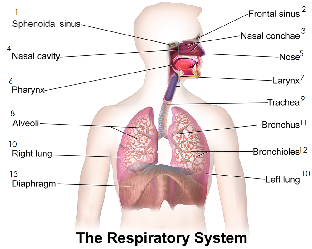
1. Sphenoidal Sinus
Sephenoidal sinuses are found in the sephenoid bone located in the skull behind the nose and eyes. They are lined with mucus membranes and are cavities that are filled with air. The main functions of the sphenoidal sinuses is to warm, moisten and filter air that is breathed in. [3]
2. Frontal Sinus
The frontal sinuses are located above the orbit of each eye in the frontal bone. These pockets of space are lined with mucus membranes, and fill with mucus which drains into the nose. [4] The frontal sinus plays a big part in the noses immune defense as it produces a vast amount of mucus which traps bacteria [5]
3. Nasal Conchae
Nasal conchae are small and thin bony elements of the upper nasal cavity. They increase the surface area of the cavities which helps with rapid warming and humidification of the air as it moves through the lungs. The nasal conchae are made up of inferior, medial, superior and supreme turbinates. [6]
4. Nasal Cavity
The nasal cavity is the highest part of the respiratory tract. It stretches from the vestibule to the nasopharynx. The four main functions that the nasal cavity has to carry out are: to heat and humidify the air that we breathe in, to extract pathogens from the air that we breathe in, to enable us to smell and also is responsible for draining the paranasal sinuses and lacrimal ducts. [7]
5. Nose
The nose is responsible for our sense of smell and is also important in the respiratory system. The nose breathes in air as well as the mouth. Small hairs inside the nose remove particles that are not needed in the body from the air. [8]
6. Pharynx
The Pharynx is a hallway, in a sense that connects the mouth and nose to the Oesophagus. It is a muscular tube that has mucus membranes lining the length of it. It plays a role in both the digestive system as well as the respiratory system. The superior part of the pharynx is a tube that connects the nasal cavity to an area behind the soft palate of the mouth. The medial section is where both food and air pass through, ending just below the tongue and the inferior section is design for only food. [9]
7. Larynx
The larynx is also commonly known as the voice box. This is because the vocal chords are held here. It's function is to produce voice and aid us in breathing and swallowing. Air travels through the larynx each time we breathe. Air from the lungs passes through causing the vocal chords to vibrate. These vibrations are then controlled by the tongue, lips and palate which enables us to speak. The larynx is made up of Muscle and cartilage which are linked by elastic tissue and is situated between the pharynx and trachea. [10]
8. Alveoli
The alveoli are little sacs of air which are found in the lungs at the end of the alveolar ducts. Alveoli are grouped together as seen in the diagram below rather than separate sacs of air. It is here where gas exchange of Oxygen and Carbon Dioxide takes place. When we breathe in air the oxygen in this inspired air is diffused through the alveoli and capillary walls into a red blood cell which then carries it away around the body. As our body metabolizes it produces Carbon Dioxide which is carried in the blood to the alveoli. It diffuses out of the blood through the capillary walls and the alveoli walls where it can be breathed out. [11]
9. Trachea
The trachea is a tube that connects the larynx to the bronchi in the lungs. It's main function in the respiratory system is to provide a pathway for air to flow in and out of the lungs for respiration. The trachea starts from the base of the neck. At the alternate end the trachea splits left and right bronchi which lead to the lungs. The wall of the trachea is thin and has C shaped pieces of cartilage in the walls. Between these cartilage rings there are spaces of membranous skin (the wall of the trachea). [12]
10. Lungs
There are two lungs in the respiratory system, the left and the right. Both lungs consist of lobes, and are soft. The main job of the lungs is to extract carbon dioxide, a form of waste that is made during metabolism from the body and bring in oxygen to provide us with energy. The lungs do this by inflating as we breathe in so they can hold more oxygen and deflating as we breathe out. [13]
11. Bronchus/Bronchi
The bronchi are the two tubes that split off of the end of the trachea. These act as the main entry way for air to access the lungs. There are 3 parts to the bronchi these are: Primary bronchi, Secondary bronchi and Tertiary Bronchi. Primary bronchi are found in the top of the lungs, secondary the middle area of the lungs and tertiary at the bottom. [14]
12. Bronchioles
The Bronchioles are situated at the end of the bronchi where the bronchi start to become narrower. These thin tubes which eventually lead to air sacs called alveoli allow oxygen to be carried to all parts of the lungs. The bronchioles do not have the support from C shaped cartilage rings like the bronchi and trachea does. The main function for the bronchioles is to ensure oxygen travels successfully to the alveoli so that gas exchange of oxygen and carbon dioxide can take place. [15]
13. Diaphragm
The diaphragm is a large sheet of muscle that runs below the lungs in the lower ribs. It separates the abdominal cavity where a large portion of the digestive system is from the thoracic cavity. It is made from skeletal muscle and has the ability to contract on a volunteer basis. The job of the diaphragm is to flatten out to decrease air pressure in the thoracic cavity allowing air to flow in the lungs. [16]
The Alveoli:
[2]
1. Pulmonary Artery
The pulmonary artery carries deoxygenated blood into the alveoli so oxygen can diffuse from the alveoli into the blood which can then be carried around the body where it can be used as energy. [18]
2. Pulmonary Vein
The pulmonary vein carries oxygenated blood from the alveoli back into the body so the oxygen can be used for energy. [18]
3. Alveoli
The alveoli are sacs of air in the lungs (found at the end of the bronchioles) where gas exchange occurs. Oxygen that is breathed in is diffused into the blood to be used as energy and carbon dioxide is diffused from capillaries which can be seen in the diagram (the green veins around the alveoli) into the alveoli where it travels back into the lungs so we can breathe it out. [18]
4. Alveolar Sacs
Alveolar sacs are where the alveolar ducts open up and where the alveoli communicate with. [17]
5. Capillary Beds
The capillary beds that surround the alveoli provide a rich blood supply, the thinness of them also allows quick gas exchange. [18]
6. Bronchioles
The Bronchioles are situated at the end of the bronchi where the bronchi start to become narrower. These thin tubes which eventually lead to air sacs called alveoli allow oxygen to be carried to all parts of the lungs. The bronchioles do not have the support from C shaped cartilage rings like the bronchi and trachea does. The main function for the bronchioles is to ensure oxygen travels successfully to the alveoli so that gas exchange of oxygen and carbon dioxide can take place. [15]
Structure and how it allows efficient gas exchange:
The alveoli is made up of squamous epithelium tissue. This results in a thin surface membrane. Having a thin surface membrane helps for quick and efficient gas exchange because there is a short diffusion distance. The wall of the alveoli are one cell thick so oxygen and carbon dioxide can easily pass through. The way loads of tiny sacs are joined together to create its definitive shape increases the surface area of the alveoli which heightens the amount of gas exchange possible. The alveoli also has a very rich supply of deoxygenated blood, which means that there will never be a lack of blood for the oxygen that travels into the alveoli, to diffuse into. Also there are a high number of capillaries that surround the alveoli. These capillaries are very thin and are able to bend the red blood cells so they are in contact with the alveoli. The ability to do this and the fact that they are very thin decreases the distance that the oxygen and carbon dioxide have to diffuse through which means the rate of gas exchange will be fast. The squamous epithelium that makes up the wall of the alveoli secretes a substance call a sufactant. This sufactant helps keep the shape of the alveoli and stops it collapsing, the sufactant also helps the diffusion of the oxygen because it lets the oxygen dissolve into it so it can reach the wall of the alveoli and diffuse through into the capillary. [18]
