Assignment 1: The Digestive System
TASK 1
the digestive system is a number of organs all connected throughout the body that work together to break down the food we consume and turn it into the energy and nutrients we need to survive. The food we consume passes from the mouth through a long tube called the Gastrointestinal Tract. This tube consists of the Pharynx, oral cavity, oesophagus, and stomach and the small and large intestines. There are other organs that are important in digestion besides these though too. Food doesn't directly pass through them though; these are the liver, gallbladder and the pancreas alongside teeth, tongue and salivary glands. There are six main functions that occur within the digestive system to help and ensure nutrients and energy are extracted for use within the body:
Ingestion
Secretion
Mixing and Movement
Digestion
Absorption
Excretion
[1]
[2]
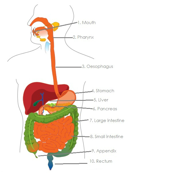
1. Mouth
The mouth is the first part of the digestive system. It contains the accessory organs teeth, tongue and salivary glands to help start the digestion of food. The teeth chop and grind up the food into small pieces suitable to swallow. The saliva then moistens the food before the tongue and a few other muscles start to push the food towards the Pharynx.
Teeth are small hard organs found around the edge of the inside of our mouths; they are made of a material called dentin with a layer of enamel over them. Inside the teeth, beneath the dentin and enamel there is a soft area known as the pulp. This area holds the blood vessels and nerves. The structure and strength of the teeth makes them perfectly designed to grind and chop up our food.
The tongue is an organ made up of several pairs of muscles. These muscles are covered in a thin, bumpy layer of skin. The outside of the tongue is covered in rough papillae which are used to grip food as the tongue moves it. It also has taste buds on the tongue that are connected to nerves; the taste buds detect taste molecules within the food and send this information to the brain via their connected nerves. Also the tongue uses its muscles to push the food toward the back of the mouth (pharynx) for further digestion.
Within our mouths we have 3 sets of salivary glands. These glands produce and secrete saliva into the mouth. The saliva wets the food and starts to break down carbohydrates. Saliva also acts as a lubricant for the food as it is passing through the mouth, pharynx and oesophagus.
2. Pharynx
the Pharynx is more commonly known as the throat. It is a funnel shaped tube connecting the mouth to the oesophagus. It acts as a bridge between the mouth and oesophagus and is responsible for transporting food that has been chewed up by the mouth to the oesophagus. However, the Pharynx is important to the respiratory system also and since it is important in two different bodily functions it has a flap of tissue called the epiglottis which helps make sure food is sent to the stomach and the air for respiration is sent to the larynx.
3. Oesophagus
The oesophagus is part of the upper gastrointestinal tract. It is a long muscular tube joining up the Pharynx to the stomach. Its purpose is to carry food from the mouth and pharynx to the stomach to be digested further. At the end of the oesophagus there is a muscle called the lower oesophageal sphincter. It is a muscular ring that is designed to close up the oesophagus after the food has passed into the stomach to trap it there.
4. Stomach
the stomach is a sac filled with stomach acid situated on the left side of the body below the diaphragm. The stomach is a major organ and acts as a place of storage for food so the body has time to digest large meals. The stomach contains Hydrochloric acid and digestive enzymes which break up food and continue the process of digestion. The Acid in the stomach also neutralizes food in our stomach. It also churns the food that is in the stomach to help further digestion, alongside this gastric juices are secreted through the walls of the stomach and mixed with the food to help break them down. The food is usually stored here for several hours.
5. Liver
The liver is an accessory organ which is situated to the right of the stomach, below the diaphragm and above the small intestine. The liver has a number of functions in the body but its main one is to create bile and to secrete it into the small intestine. It does this through bile ducts that run straight to the small intestine and the bile drains through after being produced. It also works to neutralize and extract toxins from the body to protect the body and small intestine.
Located near the liver is the Gallbladder. The gallbladders main function is to store and recycle and excess bile from the small intestine. The gallbladder can create stones which block bile ducts and stop pancreatic juices from getting through.
6. Pancreas
The pancreas is a gland that is found below the stomach. One side is connected to the duodenum and the other points to the left of the abdominal cavity. The pancreas' main job is to secrete digestive juices into the small intestine with the main aim of finishing chemical digestion of the food. The pancreatic juices (digestive juices) are full of enzymes which help break down all the food in the small intestine.
7. Large Intestine
The large intestine is a large thick tube that wraps around the border of the small intestine. The large intestines main function is to absorb water from the food and contains symbiotic bacteria that are crucial in breaking down waste so that the last small amount of nutrients left in the food can be extracted. Waste is stored here and exits the large intestine and the body via the anal canal.
8. Small Intestine
The small intestine is a long tube that is part of the lower gastrointestinal tract. It takes up the majority of space in the abdominal cavity. It is coiled up and has lots of ridges and folds that increase its surface area. This allows food to be digested better and maximizes the absorption of essential nutrients from the food. When the food leaves the small intestine about 90% of all nutrients have been extracted and absorbed from the food. [1]
9. Appendix
The function of the appendix is unknown. One theory is that is stores bacteria that are used for helping the digestive system heal after illness. Others believe it is a useless organ left over from evolution. There are no observed health problems that occur after removing the appendix. [3]
10. Rectum
the rectum is a chamber connecting the Large intestine to the anus. It is the rectums main function to remove waste from the large intestine. When the rectum needs to dispose of the waste a message is sent to the brain which then decides whether the waste can be released. If it can sphincters relax and the rectum contracts and the waste is released from the body. If it can't release the waste, the sphincters contract and the rectum holds the waste for a temporarily basis. [4]
TASK 2
The Identification of Carbohydrates (Glucose & Starch), Proteins, Lipids in different foods.
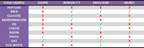
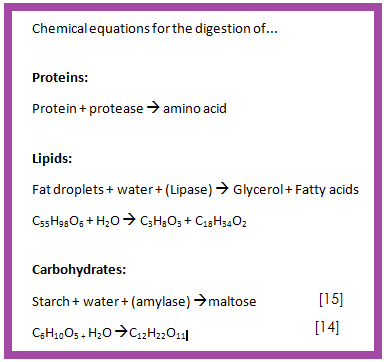
AIM: The aim of this investigation was to identify whether Carbohydrates. Proteins and Lipids were present in different foods. We used four different food tests to identify this. We used the four tests Iodine, Benedict’s reagent, Emulsion and Biuret. We used the Iodine test to identify Starches and Benedict’s to identify sugars; these were the two tests we used to identify which foods had carbohydrates in them. To test for Proteins we used the Biuret test and the Emulsion test was used to look for lipids. The foods that we tested on were peptone, milk, glucose, marshmallows, oil, cheese, raisin, pasta, oats and egg whites.
INTRODUCTION
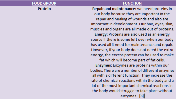
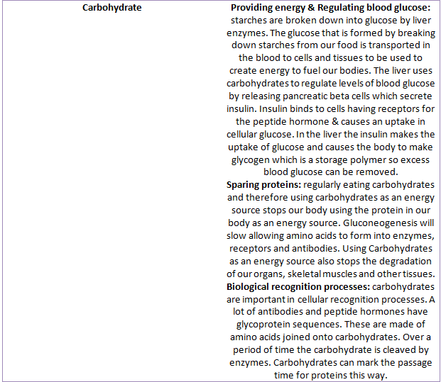
Carbohydrates, Lipids and Proteins are the three main food groups that our bodies need to survive and function properly. Carbohydrates are sugars and starches. Our body uses the carbohydrates that we consume from our food as an energy source after they have been extracted from the food during digestion. The reason they are such an ideal energy source for our body is because they can easily by broken down and changed into glucose, which is the main form of sugar that is used in the body. There are two types of carbohydrates we consume; Complex carbohydrates which are known as starches and simple which are known as sugars. Lipids are better known as fats. They have a lot of important functions within the body. These functions range from insulating your body and also protecting delicate organs, providing energy from the production of hormones and also helps in the digestion and absorption of food. They also store energy that is used to fuel the body between meals and during exercise. Any excess calories that are consumed in a day are usually stored in the form of lipids in adipose cells until they are needed. We get a lot of the lipids we need in our body from our diet. And finally Proteins are made up of amino acids and are crucial to our body. They are really important in the structure and function of the body's tissues and cells. The function of the protein is dependent on which of the 20 different amino acids it is made up of and what it's functions are. the majority of protein we get from our diet will be from eating meats as they are high in proteins. To test which foods have a high amount of carbohydrates, lipids or proteins we can do a number of different tests which will clearly show what is present in each foods. Iodine can be used to find starch, Benedict's reagent can be used to find sugars, Emulsion test can be used for lipids and Biuret reagent can be used to find proteins. [5][6][7]
METHODS
Iodine: In this experiment we put a sample of food in correctly labelled test tubes. Then to each food sample we added a few drops of Iodine. If the result was a blue/black colour the test was positive for starch.
Benedict’s reagent: A fresh sample of the foods was added to clean test tubes. Then to each test tube Benedict’s reagent and HCL was added. The test tubes were then placed in a hot water bath for around 5 minutes to heat. After the five minutes was up the test tubes were removed from the bath and the colour changes were observed. If the mixture had turned a brick red colour the test was positive for sugars.
[5]
Biuret: We added the food sample to clean test tubes that were labelled correctly. Then we added the Biuret reagent to the food samples. The test tubes were then shook and mixed well. We left each sample to stand for a few minutes and then observed any colour changes. If the sample turned violet/lilac in colour the test was positive for proteins.
Emulsion: We added our food samples to clean test tubes that were labelled up correctly. We added ethanol to each test tube and shook the test tube to mix them together. We then added a small amount of deionised water to the mixture. If a layer of cloudy white suspension forms the test is positive for lipids.
RESULTS
1. Mouth
The mouth is the first part of the digestive system. It contains the accessory organs teeth, tongue and salivary glands to help start the digestion of food. The teeth chop and grind up the food into small pieces suitable to swallow. The saliva then moistens the food before the tongue and a few other muscles start to push the food towards the Pharynx.
Teeth are small hard organs found around the edge of the inside of our mouths; they are made of a material called dentin with a layer of enamel over them. Inside the teeth, beneath the dentin and enamel there is a soft area known as the pulp. This area holds the blood vessels and nerves. The structure and strength of the teeth makes them perfectly designed to grind and chop up our food.
The tongue is an organ made up of several pairs of muscles. These muscles are covered in a thin, bumpy layer of skin. The outside of the tongue is covered in rough papillae which are used to grip food as the tongue moves it. It also has taste buds on the tongue that are connected to nerves; the taste buds detect taste molecules within the food and send this information to the brain via their connected nerves. Also the tongue uses its muscles to push the food toward the back of the mouth (pharynx) for further digestion.
Within our mouths we have 3 sets of salivary glands. These glands produce and secrete saliva into the mouth. The saliva wets the food and starts to break down carbohydrates. Saliva also acts as a lubricant for the food as it is passing through the mouth, pharynx and oesophagus.
2. Pharynx
the Pharynx is more commonly known as the throat. It is a funnel shaped tube connecting the mouth to the oesophagus. It acts as a bridge between the mouth and oesophagus and is responsible for transporting food that has been chewed up by the mouth to the oesophagus. However, the Pharynx is important to the respiratory system also and since it is important in two different bodily functions it has a flap of tissue called the epiglottis which helps make sure food is sent to the stomach and the air for respiration is sent to the larynx.
3. Oesophagus
The oesophagus is part of the upper gastrointestinal tract. It is a long muscular tube joining up the Pharynx to the stomach. Its purpose is to carry food from the mouth and pharynx to the stomach to be digested further. At the end of the oesophagus there is a muscle called the lower oesophageal sphincter. It is a muscular ring that is designed to close up the oesophagus after the food has passed into the stomach to trap it there.
4. Stomach
the stomach is a sac filled with stomach acid situated on the left side of the body below the diaphragm. The stomach is a major organ and acts as a place of storage for food so the body has time to digest large meals. The stomach contains Hydrochloric acid and digestive enzymes which break up food and continue the process of digestion. The Acid in the stomach also neutralizes food in our stomach. It also churns the food that is in the stomach to help further digestion, alongside this gastric juices are secreted through the walls of the stomach and mixed with the food to help break them down. The food is usually stored here for several hours.
5. Liver
The liver is an accessory organ which is situated to the right of the stomach, below the diaphragm and above the small intestine. The liver has a number of functions in the body but its main one is to create bile and to secrete it into the small intestine. It does this through bile ducts that run straight to the small intestine and the bile drains through after being produced. It also works to neutralize and extract toxins from the body to protect the body and small intestine.
Located near the liver is the Gallbladder. The gallbladders main function is to store and recycle and excess bile from the small intestine. The gallbladder can create stones which block bile ducts and stop pancreatic juices from getting through.
6. Pancreas
The pancreas is a gland that is found below the stomach. One side is connected to the duodenum and the other points to the left of the abdominal cavity. The pancreas' main job is to secrete digestive juices into the small intestine with the main aim of finishing chemical digestion of the food. The pancreatic juices (digestive juices) are full of enzymes which help break down all the food in the small intestine.
7. Large Intestine
The large intestine is a large thick tube that wraps around the border of the small intestine. The large intestines main function is to absorb water from the food and contains symbiotic bacteria that are crucial in breaking down waste so that the last small amount of nutrients left in the food can be extracted. Waste is stored here and exits the large intestine and the body via the anal canal.
8. Small Intestine
The small intestine is a long tube that is part of the lower gastrointestinal tract. It takes up the majority of space in the abdominal cavity. It is coiled up and has lots of ridges and folds that increase its surface area. This allows food to be digested better and maximizes the absorption of essential nutrients from the food. When the food leaves the small intestine about 90% of all nutrients have been extracted and absorbed from the food. [1]
9. Appendix
The function of the appendix is unknown. One theory is that is stores bacteria that are used for helping the digestive system heal after illness. Others believe it is a useless organ left over from evolution. There are no observed health problems that occur after removing the appendix. [3]
10. Rectum
the rectum is a chamber connecting the Large intestine to the anus. It is the rectums main function to remove waste from the large intestine. When the rectum needs to dispose of the waste a message is sent to the brain which then decides whether the waste can be released. If it can sphincters relax and the rectum contracts and the waste is released from the body. If it can't release the waste, the sphincters contract and the rectum holds the waste for a temporarily basis. [4]
TASK 2
The Identification of Carbohydrates (Glucose & Starch), Proteins, Lipids in different foods.
AIM: The aim of this investigation was to identify whether Carbohydrates. Proteins and Lipids were present in different foods. We used four different food tests to identify this. We used the four tests Iodine, Benedict’s reagent, Emulsion and Biuret. We used the Iodine test to identify Starches and Benedict’s to identify sugars; these were the two tests we used to identify which foods had carbohydrates in them. To test for Proteins we used the Biuret test and the Emulsion test was used to look for lipids. The foods that we tested on were peptone, milk, glucose, marshmallows, oil, cheese, raisin, pasta, oats and egg whites.
INTRODUCTION
Carbohydrates, Lipids and Proteins are the three main food groups that our bodies need to survive and function properly. Carbohydrates are sugars and starches. Our body uses the carbohydrates that we consume from our food as an energy source after they have been extracted from the food during digestion. The reason they are such an ideal energy source for our body is because they can easily by broken down and changed into glucose, which is the main form of sugar that is used in the body. There are two types of carbohydrates we consume; Complex carbohydrates which are known as starches and simple which are known as sugars. Lipids are better known as fats. They have a lot of important functions within the body. These functions range from insulating your body and also protecting delicate organs, providing energy from the production of hormones and also helps in the digestion and absorption of food. They also store energy that is used to fuel the body between meals and during exercise. Any excess calories that are consumed in a day are usually stored in the form of lipids in adipose cells until they are needed. We get a lot of the lipids we need in our body from our diet. And finally Proteins are made up of amino acids and are crucial to our body. They are really important in the structure and function of the body's tissues and cells. The function of the protein is dependent on which of the 20 different amino acids it is made up of and what it's functions are. the majority of protein we get from our diet will be from eating meats as they are high in proteins. To test which foods have a high amount of carbohydrates, lipids or proteins we can do a number of different tests which will clearly show what is present in each foods. Iodine can be used to find starch, Benedict's reagent can be used to find sugars, Emulsion test can be used for lipids and Biuret reagent can be used to find proteins. [5][6][7]
METHODS
Iodine: In this experiment we put a sample of food in correctly labelled test tubes. Then to each food sample we added a few drops of Iodine. If the result was a blue/black colour the test was positive for starch.
Benedict’s reagent: A fresh sample of the foods was added to clean test tubes. Then to each test tube Benedict’s reagent and HCL was added. The test tubes were then placed in a hot water bath for around 5 minutes to heat. After the five minutes was up the test tubes were removed from the bath and the colour changes were observed. If the mixture had turned a brick red colour the test was positive for sugars.
[5]
Biuret: We added the food sample to clean test tubes that were labelled correctly. Then we added the Biuret reagent to the food samples. The test tubes were then shook and mixed well. We left each sample to stand for a few minutes and then observed any colour changes. If the sample turned violet/lilac in colour the test was positive for proteins.
Emulsion: We added our food samples to clean test tubes that were labelled up correctly. We added ethanol to each test tube and shook the test tube to mix them together. We then added a small amount of deionised water to the mixture. If a layer of cloudy white suspension forms the test is positive for lipids.
RESULTS
CONCLUSIONS
Iodine: From our results it clearly shows that the pasta and oats were the only two foods that we tested on that contained starch as they were the only ones that turned a blue/black colour when the iodine was added to them.
Benedict's reagent: The Benedict's reagent is used to look for reducing and non-reducing sugars. In our test milk, glucose, marshmallow, raisins and oats all gave positive results by turning a brick red colour at the end of the test. This shows that there are sugars present within these foods.
Emulsion: Our results show for the emulsion test that milk, oil, cheese, oats and egg whites were all positive results. This shows that these foods all contain lipids. We can tell this by the white precipitate that formed after we added the ethanol and deionised water.
Biuret: In this test our only positive results were for milk, marshmallows, raisins and egg whites. They all turned a purple/violet/lilac colour when the biuret solution was added which clearly showed that they contained proteins.
TASK 3
TASK 4
Epithelial
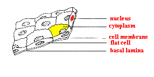
Epithelial tissues are linings on this inside and outside of the body. They are present in organs and skins. Epithelial tissues are made up of closely packed flat cells that don't contain a lot of inter cellular material. This type of tissue doesn't contain blood vessels and waste and nutrients are exchanged by diffusion from connective tissues nearby. The top surface of epithelial tissue always faces the outside of the body or an inner body cavity and the bottom layer always lies on connective tissue. A thin layer called the basement membrane forms between the epithelial tissue and the connective tissue. [16]
Epithelial tissue is formed by layers of tightly packed cells piled on top of each other. [17]
Connective
Connective tissue supports and joins other tissues in the body. The way connective tissue is structured is it has cells spread through an extra cellular matrix of fibrous proteins. It also has a basement membrane that has glycoproteins attached to it. Connective tissue is important to us as it forms a mechanical structure or frame work known as the skeleton which enables us to move. It has a large amount of inter cellular space and inter cellular material and very few cells which are spaced out throughout the inter cellular space. [18]
Nervous
Nervous tissue is responsible for carrying impulses, stimuli and messages around the body. A standard part of the nervous system is a neuron. Neurons have 2 main parts to their structure; these are the cell body and the nerve processes. The cell body contains the nucleus, cytoplasm and organelles. The nerve processes are branches that are able to send and receive signals. Axons are nerve processes carry signals away from the cell body and Dendrites carry signals towards the cell body. [19]
Nervous tissue is responsible for carrying impulses, stimuli and messages around the body. A standard part of the nervous system is a neuron. Neurons have 2 main parts to their structure; these are the cell body and the nerve processes. The cell body contains the nucleus, cytoplasm and organelles. The nerve processes are branches that are able to send and receive signals. Axons are nerve processes carry signals away from the cell body and Dendrites carry signals towards the cell body. [19]
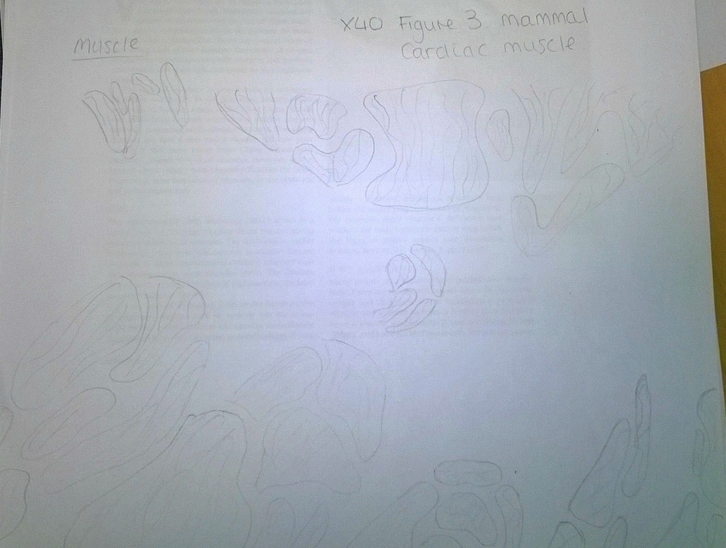
Muscle
Muscle tissue is made up of cells that are able to contract. They contain a number of micro filaments that are made of contractile proteins which are called actin and myosin. [20] There are three different types of muscle tissues that all have slightly different structures. These three are Smooth Muscle Tissue, Skeletal Muscle Tissue and Cardiac Muscle Tissue. Smooth muscle tissue is made up of long and thin muscle cells fibres. At the end of the fibres there is a nucleus. The cells are filled with a cytoplasm which is surrounded by a cell membrane. Smooth muscle fibres intertwine to form sheets and layers. Skeletal muscles are attached to various bones in the skeleton and enable them to move. The muscle tends to be covered in connective tissue. These connective tissue then folds around small muscular bundles called fasciculi. These consist of small cylinder shaped muscles that contain nuclei. The sarcoplasm has light and dark bands that give the cell a striated appearance. Cardiac muscle tissue is only found in the heart it has characteristics of both skeletal and smooth muscle tissues. Its fibres are striated and have a lot of nuclei like skeletal however, it is involuntary muscle like smooth muscle tissue is. It differs from striated as its striations are not as obvious, they're shorter and the sarcolemma is thinner. It only has one nucleus in each cardiac fibre, they are branched and the branches are then connected to each other by muscle bridges. [21]
Muscle tissue is made up of cells that are able to contract. They contain a number of micro filaments that are made of contractile proteins which are called actin and myosin. [20] There are three different types of muscle tissues that all have slightly different structures. These three are Smooth Muscle Tissue, Skeletal Muscle Tissue and Cardiac Muscle Tissue. Smooth muscle tissue is made up of long and thin muscle cells fibres. At the end of the fibres there is a nucleus. The cells are filled with a cytoplasm which is surrounded by a cell membrane. Smooth muscle fibres intertwine to form sheets and layers. Skeletal muscles are attached to various bones in the skeleton and enable them to move. The muscle tends to be covered in connective tissue. These connective tissue then folds around small muscular bundles called fasciculi. These consist of small cylinder shaped muscles that contain nuclei. The sarcoplasm has light and dark bands that give the cell a striated appearance. Cardiac muscle tissue is only found in the heart it has characteristics of both skeletal and smooth muscle tissues. Its fibres are striated and have a lot of nuclei like skeletal however, it is involuntary muscle like smooth muscle tissue is. It differs from striated as its striations are not as obvious, they're shorter and the sarcolemma is thinner. It only has one nucleus in each cardiac fibre, they are branched and the branches are then connected to each other by muscle bridges. [21]
TASK 5
The human body is made up of different types of cells, tissues and organ systems that all contribute and carry out crucial functions within the body which enables us to live. There are four main types of tissues that make up all that we are; these are epithelial, connective, muscle and nervous.
Epithelial tissue covers the body, outside and in. It is formed by cells that are closely packed together and are layered over each other. Epithelial is specialized to act as a lining for any surface in the body. The type of epithelial tissue that is found on internal surfaces is known as endothelium. Separating the epithelium tissue from any underlying tissue by a thin strip of connective tissue which is known as the There are two types of epithelium tissue simple epithelium which is just one cell thick and stratified epithelium which is two or more cells thick. There are a number of different types of Simple Epithelium cells. The first is Squamous epithelium: squamous epithelium cells are flat in shape and are very thin. This type of epithelium cells are found in the linings of the mouth, blood vessels and our hearts and lungs. They also make up the outside layers of our skin.
Simple cuboidal epithelium cells are cuboidal in shape; each cell has a circular nucleus in the cell centre. This type of epithelium cell and tissue can be found within glands and gland ducts and the linings of the kidney tubules.
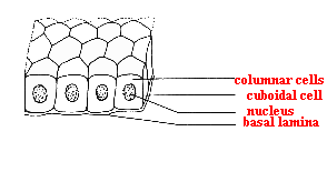
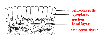
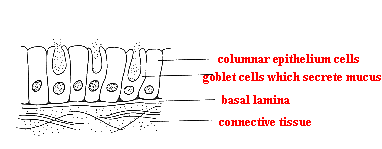
Simple columnar epithelium are elongated and shaped like columns; the nucleus of a simple columnar epithelium is usually found at the base of a cell and is elongated in shape. These types of cells form the tissue that lines the stomach and intestines. There are also columnar epithelium cells that have become specialized in such a way that enables them for sensory reception. These types of specialized cells are found in the nose, ears and also the taste buds on your tongue. Also between columnar epithelium cells that line the duodenum cells known as goblet cells are found. These cells have the ability to secrete mucus which lubricates the surface and keeps it smooth.
[22]
Glandular epithelium cells are specialized epithelium cells that have the ability to secrete substances in the body; it is this type of cell that when they form tissue makes glands. There are two types of glands glandular epithelium cells can make. These are endocrine and exocrine. These glands then secrete substances such as enzymes, hormones and saliva. A good example of when glandular epithelium cells are found is in the saliva gland which starts the breakdown of foods in the mouth. [23] [22]
Glandular epithelium cells are specialized epithelium cells that have the ability to secrete substances in the body; it is this type of cell that when they form tissue makes glands. There are two types of glands glandular epithelium cells can make. These are endocrine and exocrine. These glands then secrete substances such as enzymes, hormones and saliva. A good example of when glandular epithelium cells are found is in the saliva gland which starts the breakdown of foods in the mouth. [23] [22]
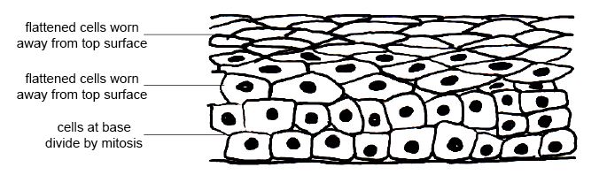
Stratified epithelium tissue is a more complex type of epithelium as it is made up of lots of layers of epithelium cells. This type of tissue are found in areas of the body that undergo a lot of wear and tear. The cells on the top layer of stratified epithelium are usually flat and scaly and sometimes contain keratin. An example of where stratified epithelium tissue is found is in the lining of the mouth but it does not contain keratin. [22]
Connective tissues main role in the body is to support, connect and bind the different tissues of the body together. It is also what provides the framework that enables our bodies to move; the skeleton. Connective tissue has a large amount of intercellular substance that is known as the matrix. This matrix is composed up of ground substances and fibers. In general connective tissue the ground substance is most commonly water and the fibers are usually collagen. Connective tissues have a small amount of cells that are spread out separate from each other. The few cells that are in connective substance are what are responsible for secreting the matrix; this matrix will be a non-living substance. The substance could be liquid, semi-solid or solid. An example of a liquid matrix would be blood, semi-solid would be connective tissue and solid bone. Connective tissue can be broken down into four different types; these are adipose tissue, cartilage, blood and bone. [24] [25]
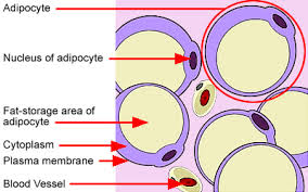
Adipose tissue is a type of connective tissue that is loose and fibrous and it is full of fat cells that are called adipocytes. These fat cells are specialized to store triglycerides. Each cell has a fat droplet inside which fills the majority of the cell. The fat pushes the nucleus and other organelles towards the edge of the cell. Adipose tissues are found in thick layers under the skin and also around the kidneys. Beside insulation and preventing heat loss this type of connective tissue especially in the case of the kidneys and other important organs that it surrounds its main function is to protect, cushion and support. It is also an means of energy storage and excess food within the body is converted to fat and stored in the cells of this tissue. [26]
Adipose tissue is a type of connective tissue that is loose and fibrous and it is full of fat cells that are called adipocytes. These fat cells are specialized to store triglycerides. Each cell has a fat droplet inside which fills the majority of the cell. The fat pushes the nucleus and other organelles towards the edge of the cell. Adipose tissues are found in thick layers under the skin and also around the kidneys. Beside insulation and preventing heat loss this type of connective tissue especially in the case of the kidneys and other important organs that it surrounds its main function is to protect, cushion and support. It is also an means of energy storage and excess food within the body is converted to fat and stored in the cells of this tissue. [26]
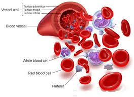
Blood is a connective tissue as it has a matrix there are two types of living cells within blood, these are erythrocytes and leukocytes and the matrix is usually called the plasma. Erythrocytes are cells that are specialized to carry oxygen around the body and also carbon dioxide out of the body. The main purpose blood has in relation to the digestive system is it supplies oxygen to the different organs that are taking part in breaking down the food giving the organs energy to function and also the plasma absorbs nutrients from digestion and carries them to the correct parts of the body that needs the specific nutrients that it has absorbed. [28]
[29]
All these different cells and tissues then forge together to make organs. Within the Digestive system there are many organs that all do their part to break down food and extract all the nutrients that we need. The Stomach is made up of epithelial tissues, Muscle tissues and Glandular tissues. The epithelial tissue covers all the surface of the outside and the inside of the stomach, the muscle tissues purpose is to churn the stomach's contents and the glandular tissues purpose is to produce digestive juices that aid in the stage of digestion taking part in the stomach. These glandular tissues produce acid and enzymes needed for digestion. The stomach is made up of different layers. These layers are called the mucosa, submucosa, muscularis and serosa.
The mucosa is the inner layer and is made of mucous membrane. This mucous membrane contains a lot of simple columnar epithelial cells and gastric pits. The gastric pits contain a large amount of exocrine cells that are responsible for the secretion of Hydrochloric acid and digestive juices into the stomach. There are mucous cells that are also located in the gastric pits and throughout the lining of the stomach which to protect the stomach from its own digestive juices secrete mucous.
The submucosa surrounds the mucosa, it is made up of a variety of connective tissues, nerves and blood vessels. The connective tissues that are in the submucosa are responsible for connecting the mucosa to the muscularis and also supporting the mucosa. The supply of blood to the submucosa carries nutrients to the wall of the stomach; and finally the nerves in the submucosa monitor what is in the stomach and also controls the contraction of the muscles to make sure they are smooth. The nerves also control and regulate the secretion of digestive juices into the stomach.
The muscularis is found surrounding the submucosa and makes up the majority of the stomach's mass. The muscularis has three layers of muscle tissue that are positioned in a way that their fibers all run in a different direction. This smooth muscle allows the stomach to contract enabling the food to be mixed and moved through the digestive tract. Also situated inside the mucosa is a smooth and thin muscular layer called the muscularis mucosae which enables the mucosa to fold and increase the amount of contact it has with the stomach's contents.
The serosa is the most immediate outside layer of the stomach. It is a serous membrane which is quite thin. It is made up of simple squamous epithelial tissue and a type of connective tissue called areolar connective tissue. The surface of the serosa is smooth and slippy. It secretes a watery fluid called serous fluid; its smooth surface reduces friction as the stomach enlarges after a meal or as it moves while mixing and moving the food through the digestive tract. [30]
All our organs work together in systems to keep our bodies working and functioning so we can live a healthy life. The mouth chews up and starts the breakdown of food and then saliva glands moisten the food so it can pass down the oesophagus to the stomach where the digestion is taken further. Also all our different organ systems within our bodies are all connected in ways. Say if the cardiovascular system wasn't working or was removed from the body the Respiratory system would suffer as the blood wouldn't be circulating around the body which means there would be a lack of Oxygen being transported around the body. This would result in our cells dying.
Our bodies would fail to work without all of our organs and how the tissues and cells all interlock in certain ways that create a perfect system that enables us to live a healthy life. It is amazing how all our organs and systems are made in such a way that the majority of what our bodies need to do to live is done by our bodies themselves without us really thinking about it. This is down to the specialized cells and tissues that make up everything that we are.
